A working catalog across orthobiologics, regenerative therapy, spinal fixation, and surgical imaging, selected because they address different parts of the same biological cascade. Full specs, manufacturer detail, and pricing are shared directly, under agreement, once you're talking to us.
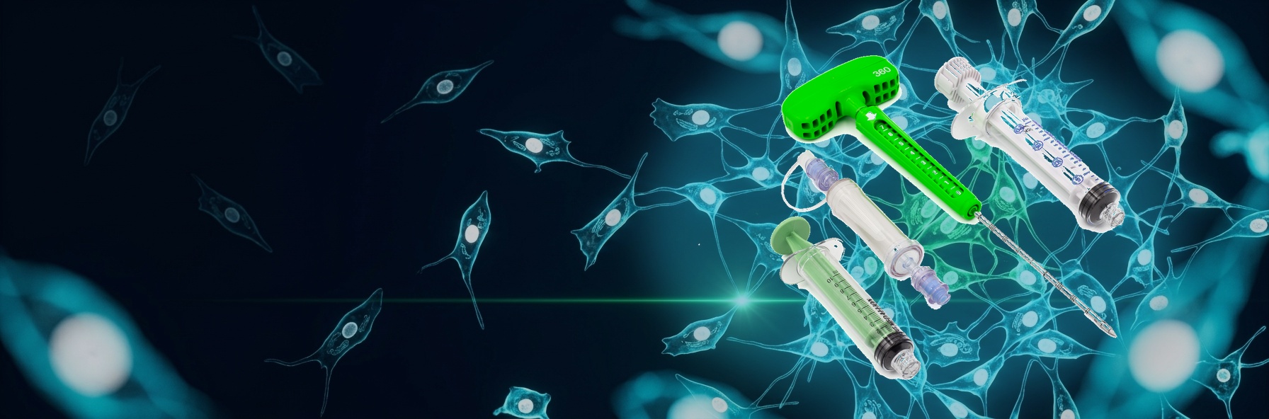
Orthobiologics are biological materials used in modern surgical practice to promote faster, more natural healing. Mesenchymal stem cell (MSC) therapy is one of the core tools in that category, harnessing the body's own regenerative capacity to repair damaged tissue. The two primary sources are bone marrow, drawn from the iliac crest where MSC concentration is highest, and adipose tissue, which can be harvested through a minimally invasive, low-risk procedure. These therapies apply across orthopaedics, spinal surgery, maxillofacial surgery, pain management, wound healing, and general or urogynecological surgery.
A continuously wearable bone growth stimulation system built to fit into a patient's day rather than interrupt it, compact enough to clip on a belt or tuck into a pocket, with no restricted postures and no scheduled sessions. Built-in sensors and a proprietary algorithm track wear time and relative activity level automatically, combined with patient-reported pain scores in a companion app for a clear day-to-day view of recovery. Clinicians get that same picture through a remote portal that flags patients whose progress may be falling off track. Preclinical data also shows increasing bone cell proliferation the longer the device is worn each day, consistent with a dose-dependent response.
The mechanism is well characterized. Work by Brighton and colleagues established that capacitively coupled signal transduction in bone cells proceeds through voltage-gated calcium channels — proliferation is blocked by verapamil, a calcium channel blocker, but not by inositol-phosphate inhibitors, which distinguishes it from the inductive-coupling pathway. The resulting rise in cytosolic calcium activates calmodulin and upregulates TGF-β1 and other osteopromotive factors.
The clinical evidence supports the modality independently of any one device. A Level I systematic review and meta-analysis of randomized controlled trials (n=941) found postoperative electrical stimulation increased the odds of successful fusion 2.5-fold relative to control, with no significant subgroup interaction by stimulation type. In a randomized, double-blind, placebo-controlled trial of capacitively coupled stimulation across 28 U.S. surgical sites, 84.7% of actively stimulated patients met the combined clinical and radiographic success criteria versus 64.9% on placebo.
A point-of-care aspiration system built around a specific premise: what matters in a marrow graft is not total cell count but the number of true early-stage stem cells, measured as colony-forming unit-fibroblast (CFU-f). In clinical work on non-union and osteonecrosis, CFU-f content was the only measured variable that reached statistical significance — not total nucleated cells, not platelets.
The device is designed to protect that fraction. A closed and sealed distal tip means 100% of the harvest is lateral — drawn perpendicular to the channel the cannula creates, rather than through an open end where peripheral blood dominates the draw. A micrometric gear retracts the cannula 5 mm with each 360° rotation, so every 0.5–1 mL pull comes from a fresh marrow site without repositioning the device. The result is many small high-quality aspirates instead of one large diluted one, consistent with published work showing that small-volume, small-syringe technique yields greater CFU-f.
It is a single-step, centrifuge-free device: no processing time, no machinery loan, no dedicated staff, and no reason to leave the sterile field. The concentrate is ready to use at the point of care. Per manufacturer data, roughly 40% more cells survive than in centrifuge-based systems.
That matters because centrifuge-based concentration discards roughly 85% of the aspirate and leaves an estimated 40% of stem cells behind in the discarded red cell fraction — including dense, actively cycling anti-inflammatory progenitors that always sediment with the red cells. Centrifugation also cannot distinguish marrow-derived nucleated cells from peripheral blood ones, since both share the same density. In older patients, that concentrates inflammatory macrophages and neutrophils along with everything else.
The relevance is age-dependent. Circulating platelet and white cell counts hold up over a lifetime, but the body's ability to mobilize marrow stem cells to a site of trauma declines substantially with age. In older or healing-impaired patients, the vasculogenic signal from platelets alone is often not enough to complete the cascade. The concentrated aspirate can be combined with a bone graft substitute to carry those cells to the defect and hold them there.
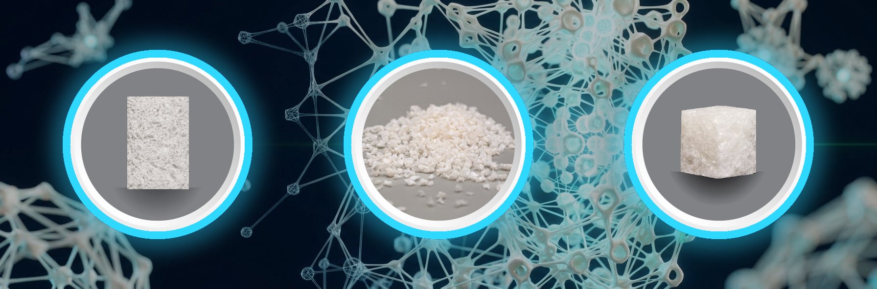
A hyper-crosslinked carbohydrate polymer originally developed as a substrate for stem cell culture and differentiation, now doing that same work inside the graft site. It optimizes the cellular microenvironment for bone formation and supports blood vessel ingrowth, so what fills the defect is vascularized, healthy bone rather than inert filler. Degradation is tuned to match the rate of regeneration, and because it starts radiolucent and turns radiopaque as fusion progresses, surgeons can watch the fusion happen instead of inferring it months later. Fully synthesized, so quality is consistent lot to lot with zero risk of disease transmission. Expands back to volume after compression, trimmable, stores at room temperature, and available in block, granulate, and cube forms — or custom shapes on request.
In a peer-reviewed retrospective study of 63 spinal fusion procedures, the polymer achieved a 91.8% overall fusion rate: 100% thoracic, 95% lumbar, and 85% cervical. Regions fused with it showed visible osseous formation within three months postoperatively and efficacy comparable to other bone graft substitutes at six months. Used as the sole bone graft substitute in a 29-patient subset, the fusion rate was 86.2%. No adverse events related to its use were observed. A separate ovine model showed bone growth equivalent to autograft on CT at six months with no histologic abnormalities. A separate retrospective cervical study reported fusion rates exceeding 90% by three months, comparable to i-Factor and autograft at 12 and 24 months. Published work also covers critical-size defect repair, where bone density at the graft site rose over sixteen weeks while autograft sites declined, and foot and ankle limb salvage.
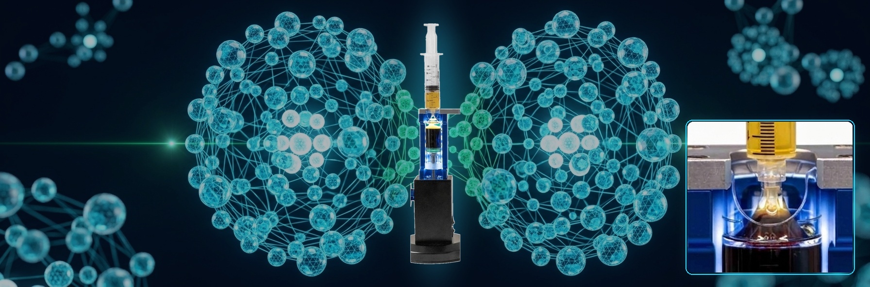
A single FDA 510(k)-cleared benchtop system that processes three distinct biologics from the patient's own blood or marrow: platelet-rich plasma, bone marrow aspirate concentrate, and protein concentrate (A2M and IL-1ra). One 10-minute spin, prepared chairside, with fewer transfer steps than most systems on the market — less mechanical trauma to cells and lower contamination risk.
Platelet-rich plasma. Captures the buffy coat through a simple push method with clear volume markings for reproducible results. Leukocyte concentration is tailorable: the volume of enriched plasma, buffy coat, and white cells can be adjusted to fit the patient and the pathology.
Protein concentrate (A2M). Alpha-2-macroglobulin works by a different mechanism than platelets. Rather than delivering growth factors, it acts as a broad-spectrum protease inhibitor — capturing and neutralizing the matrix metalloproteinases and aggrecanases that degrade cartilage, and downregulating IL-1 and NF-κB inflammatory signaling. In a rat model of osteoarthritis, supplemental intra-articular A2M was identified as a master inhibitor of cartilage-degrading factors and attenuated the progression of post-traumatic OA. For a degenerative joint, that means protecting the matrix rather than only stimulating repair.
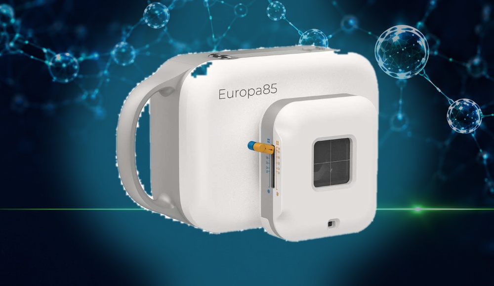
The Europa85 from Aspen Imaging Healthcare is a handheld X-ray unit weighing 7.9 lb, built for mobile teams and smaller practice settings that need flexibility without giving up image quality. Unlike conventional portable units, images are captured and reviewed directly on the device via a 7″ HD touch screen, then transmitted wirelessly to a PC through the integrated Intel workstation. Multi-leaf collimator, 0.8 mm focal spot, and battery charging from a standard 120V outlet.
GRIFFIN™ is a fully titanium expandable cage from Diagon Medical, manipulated to align in the sagittal plane and then uniquely realigned coronally by the surgeon. Delivered through a minimally invasive corridor to reduce blood loss and operative time. The graft window is where this platform converges: the cage establishes and holds the mechanical environment, and the biologic carrier packed into it recruits the cells and growth factors that turn a stable segment into a fused one.